Medullary pyramids (brainstem)
Appearance
| Medullary pyramids (brainstem) | |
|---|---|
 Medulla oblongata and pons. Anterior surface. (Pyramid labeled at center.) | |
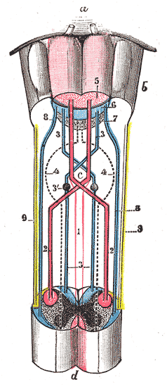
 Decussation of pyramids. Scheme showing passage of various fasciculi from medulla spinalis to medulla oblongata. a. Pons. b. Medulla oblongata. c. Decussation of the pyramids. d. Section of cervical part of medulla spinalis. 1. Anterior cerebrospinal fasciculus (in red). 2. Lateral cerebrospinal fasciculus (in red). 3. Sensory tract (fasciculi gracilis et cuneatus) (in blue). 3’. Gracile and cuneate nuclei. 4. Antero-lateral proper fasciculus (in dotted line). 5. Pyramid. 6. Lemniscus. 7. Medial longitudinal fasciculus. 8. Ventral spinocerebellar fasciculus (in blue). 9. Dorsal spinocerebellar fasciculus (in yellow). | |
| Details | |
| Identifiers | |
| Latin | pyramis medullae oblongatae |
| NeuroNames | 705 |
| TA98 | A14.1.04.003 |
| TA2 | 5985 |
| FMA | 75254 |
| Anatomical terminology | |
The anterior district of the medulla oblongata is named the pyramid and lies between the anterior median fissure and the antero-lateral sulcus.
Its upper end is slightly constricted, and between it and the pons the fibers of the abducent nerve emerge; a little below the pons it becomes enlarged and prominent, and finally tapers into the anterior funiculus of the medulla spinalis, with which, at first sight, it appears to be directly continuous.
See also
- Corticospinal tract (also known as "pyramidal tract")
- Decussation of the pyramids
External links
![]() This article incorporates text in the public domain from page 768 of the 20th edition of Gray's Anatomy (1918)
This article incorporates text in the public domain from page 768 of the 20th edition of Gray's Anatomy (1918)
